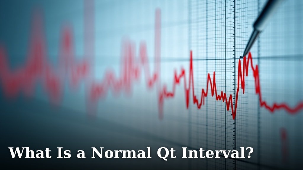
How Do You Know If Your ECG Reading Is Normal?
On this page (8 sections)
Knowing if your ECG reading is normal can feel like deciphering a secret code. Generally, a normal ECG reading shows a regular heart rhythm and electrical signals that appear as smooth, consistent waves. Your doctor will look at several key factors to determine if everything is in order, but in simple terms, it means your heart’s electrical activity looks healthy and balanced.
While a “normal” ECG can vary slightly from person to person, there are specific patterns doctors look for. They assess the heart rate, the duration of each electrical impulse, and the overall shape of the waveforms. Any significant deviations from these typical patterns might suggest an underlying heart condition that needs further investigation.
- What a normal ECG looks like: Smooth waves, regular rhythm.
- Key things doctors check: Heart rate, impulse duration, waveform shape.
- Why it matters: Detects potential heart issues early.
- Always consult your doctor: They are the best resource for interpreting your results.
Let’s dive deeper into what makes an ECG reading appear normal and what those waves and lines actually mean. We’ll help you understand the basics so you can have a more informed conversation with your healthcare provider.
Understanding Your ECG: What Does a “Normal” Look Like?
So, you’ve had an electrocardiogram (ECG), and now you’re wondering what those squiggly lines mean. It’s totally normal to feel a bit in the dark! Think of an ECG as a snapshot of your heart’s electrical activity. A normal reading shows that your heart is beating in a regular rhythm and at a healthy pace. It means the electrical signals are traveling through your heart muscle as they should, causing it to pump blood efficiently.
The key takeaway is that a normal ECG suggests your heart’s electrical system is working well. It doesn’t necessarily mean there are no heart issues, but it’s a very good sign that the electrical plumbing is functioning correctly. Your doctor uses this as one piece of the puzzle to assess your overall heart health.
The Anatomy of a Normal ECG Waveform
An ECG traces the electrical journey of your heartbeat. This journey creates a unique pattern of waves. These waves have names: P wave, QRS complex, and T wave. Each one represents a different part of the heart’s pumping cycle. When these waves appear in a consistent, predictable pattern, it’s a good indicator of a normal ECG.
The P Wave: Setting the Stage
The P wave is the very first little bump you’ll see. It represents the electrical impulse originating in the upper chambers of your heart, called the atria. This impulse causes the atria to contract, pushing blood down into the lower chambers, the ventricles. A normal P wave is typically smooth and rounded. Its presence and shape tell your doctor that the atria are being stimulated electrically.
The QRS Complex: The Main Event
Following the P wave is the QRS complex. This is usually the largest and most prominent part of the ECG tracing. It represents the electrical impulse spreading through the ventricles. When this signal fires, it causes the ventricles to contract forcefully, pumping blood out to your lungs and the rest of your body. A normal QRS complex is sharp and well-defined. Its size, shape, and timing are crucial for determining if the ventricles are working correctly (Cleveland Clinic).
The T Wave: The Heart’s Reset
After the QRS complex, you’ll see the T wave. This wave represents the electrical recovery of the ventricles. It’s like the heart muscle getting ready for the next beat. A normal T wave is usually smooth and slightly asymmetrical. Its shape and direction can give your doctor clues about the overall health of the heart muscle and electrolyte balance in your body.
What Doctors Look for Beyond the Waves
While understanding the waves is helpful, your doctor looks at several other factors on the ECG to make a diagnosis. They aren’t just looking at individual waves; they’re examining the entire pattern and rhythm. It’s like looking at a whole musical score, not just one note.
Heart Rate: The Speed of Your Beat
One of the first things assessed is your heart rate. For adults at rest, a normal heart rate typically falls between 60 and 100 beats per minute. If your heart rate is consistently too slow (bradycardia) or too fast (tachycardia), it might indicate an issue. Your doctor will consider your activity level and any medications you’re taking, as these can affect your heart rate.
Heart Rhythm: The Beat’s Consistency
Beyond just the rate, the regularity of your heartbeat is key. A normal ECG shows a regular rhythm, meaning the time between each heartbeat is consistent. Irregular heartbeats, known as arrhythmias, can range from minor skips to more serious disruptions. The ECG helps identify patterns that suggest an irregular rhythm.
Electrical Axis: The Heart’s Direction
This might sound a bit technical, but the electrical axis refers to the general direction of the electrical current in your heart. In a healthy heart, this axis falls within a specific range. Deviations can sometimes point to issues like an enlarged heart chamber or a previous heart attack (NIH).
What Makes an ECG Reading “Abnormal”?
An “abnormal” ECG doesn’t always mean something is seriously wrong, but it does mean there’s a deviation from the expected pattern. It’s a signal that further investigation might be needed. These deviations can include changes in the shape, size, or timing of the waves, or issues with the heart rate and rhythm, which may help explain inconsistent ECG readings.
Irregular Rhythms (Arrhythmias)
As mentioned, irregular rhythms are a common finding. Conditions like atrial fibrillation (AFib), where the upper chambers quiver irregularly, or premature ventricular contractions (PVCs), where the ventricles beat too early, will show distinct patterns on the ECG. These need careful evaluation by your doctor. Irregular rhythms like AFib or PVCs will show distinct patterns on the ECG; our guide explains how an ECG can detect ectopic beats.
Signs of Heart Muscle Damage or Strain
Changes in the QRS complex or the ST segment (the flat line between the QRS and T wave) can sometimes indicate that the heart muscle isn’t getting enough oxygen. This could be a sign of angina or a myocardial infarction (heart attack). Certain abnormalities might suggest previous damage too.
Electrolyte Imbalances
Your heart’s electrical activity relies on the right balance of electrolytes like potassium and calcium in your body. If these levels are too high or too low, it can affect the appearance of the T wave and other parts of the ECG. This is why doctors often check your blood work alongside your ECG.
When to Discuss Your ECG Results with Your Doctor
It’s important to remember that you shouldn’t try to self-diagnose based on your ECG. While understanding the basics is empowering, only a qualified healthcare professional can interpret the results in the context of your overall health. If your doctor mentions any of the following, it’s definitely worth having a detailed conversation:
- An unusual heart rate (too fast or too slow).
- An irregular heartbeat or skipped beats.
- Any mention of “arrhythmia” or specific rhythm abnormalities.
- Changes in the ST segment or T waves.
- Anything that sounds like it deviates from “normal.”
Your doctor will consider your symptoms, medical history, and other tests. The ECG is just one tool in their diagnostic toolkit. They will explain what the findings mean for you personally and what, if any, next steps are recommended.
Key Things to Remember About Your ECG
Navigating your health information can sometimes feel overwhelming. Here’s a quick rundown of the most important points about your ECG reading:
- A normal ECG shows a regular rhythm and consistent wave patterns.
- Doctors check heart rate, rhythm regularity, and wave characteristics.
- Waves like P, QRS, and T represent different stages of your heartbeat.
- Deviations can signal various heart conditions, but require professional interpretation.
- Always discuss your results with your healthcare provider for accurate understanding.
Think of your ECG as a report card for your heart’s electrical system. A good report card is reassuring, and a less-than-perfect one is an invitation to work with your doctor to keep your heart as healthy as possible!
Conclusion
Understanding your ECG reading is a powerful step toward managing your heart health. You’ve learned that a normal ECG shows a steady rhythm and predictable wave patterns, reflecting your heart’s efficient electrical function. It’s reassuring when your ECG looks “normal,” but remember, it’s just one part of your doctor’s assessment. If your results show any deviations, it’s an opportunity for your doctor to investigate further and ensure you receive the best care. Don’t hesitate to ask questions and partner with your healthcare provider. Your next step is to schedule a follow-up appointment with your doctor to discuss your specific ECG results and any concerns you might have.
Frequently Asked Questions
Can I interpret my ECG results myself?
It’s generally not recommended to interpret your ECG results on your own. While understanding the basics of waves and rhythms is helpful, a qualified healthcare professional considers your entire medical history and symptoms. They are best equipped to accurately diagnose any potential issues shown on the ECG. For a look at how software reads ECG signals, see our guide to AI analysis of ECGs.
What if my ECG is normal, but I still have symptoms like chest pain?
A normal ECG is a good sign, but it doesn’t rule out all heart problems. Some heart conditions might not be evident on a resting ECG. If you’re experiencing symptoms like chest pain, it’s crucial to discuss this with your doctor. They may recommend further tests to get a complete picture of your heart health.
How often should I have an ECG done?
The frequency of ECGs depends on your individual health status and risk factors. If you have a history of heart problems or certain risk factors, your doctor might recommend regular ECGs. For healthy individuals with no known issues, ECGs are typically done as needed during check-ups or if symptoms arise.
What does it mean if my ECG shows a slightly fast or slow heart rate?
A heart rate outside the typical 60-100 beats per minute range can mean several things. It might be due to factors like exercise, stress, medications, or an underlying medical condition. Your doctor will evaluate whether your heart rate is consistently too fast (tachycardia) or too slow (bradycardia) and determine if further investigation is needed.
Can stress or anxiety affect my ECG reading?
Yes, significant stress or anxiety can sometimes influence your heart rate and rhythm, which might be reflected on an ECG. Your doctor understands that temporary factors can affect readings and will consider your overall situation. If you notice your ECG changes during periods of high stress, it’s worth mentioning to your doctor.