How Does an ECG Monitor Detect AFib?
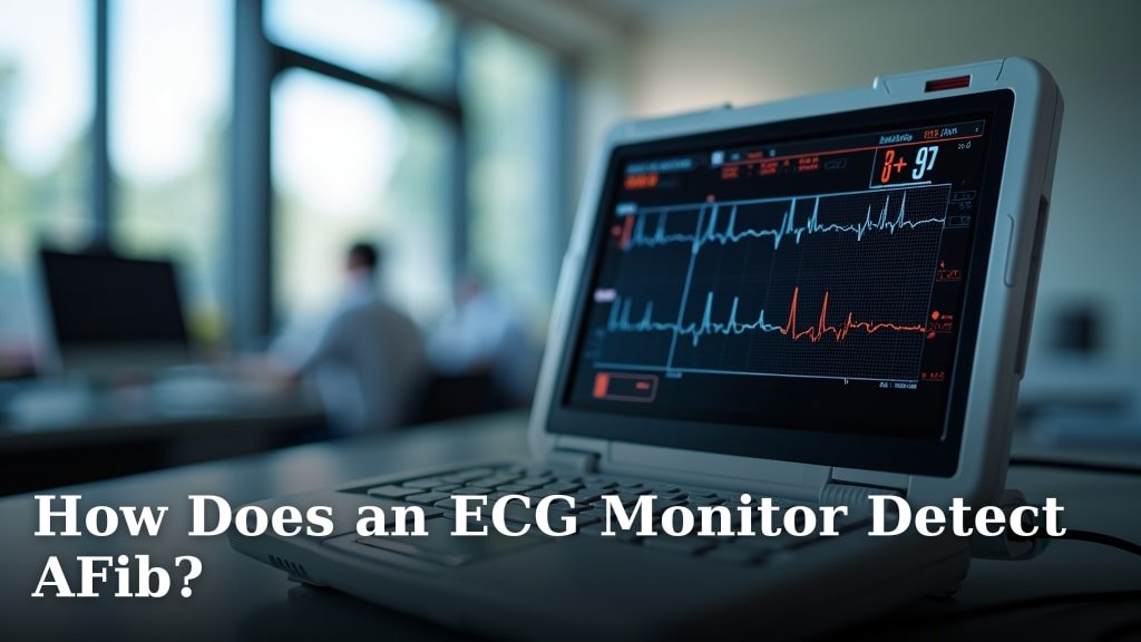
An ECG monitor detects AFib (Atrial Fibrillation) by recording the electrical signals of your heart. It looks for irregular patterns, especially chaotic electrical activity in the upper chambers of your heart, which is a hallmark of AFib.
You might be wondering how a small device can tell if your heart is beating unusually. We found that these monitors capture your heart’s rhythm and then analyze it to spot the disorganized electrical impulses that indicate AFib.
- ECG monitors record heart’s electrical signals.
- They identify irregular and chaotic heart rhythms.
- AFib causes disorganized electrical activity in the atria.
- These devices help diagnose AFib early.
Ready to understand the nitty-gritty? Let’s walk through exactly how an ECG monitor helps identify this common heart condition, step by step.
How an ECG Monitor Pinpoints Atrial Fibrillation
You’re curious about how these clever devices work, right? We found that an ECG monitor doesn’t just listen to your heartbeat; it interprets the electrical language of your heart. Think of it like reading a secret code that only your heart knows.
When your heart is healthy, these electrical signals follow a predictable, rhythmic path. With AFib, that path becomes chaotic and disorganized. The ECG monitor is designed to spot these specific deviations.
The Heart’s Electrical Symphony
Your heart is a remarkable pump, and its rhythm is controlled by tiny electrical impulses. These impulses tell different parts of your heart when to contract. It’s like a well-orchestrated band, with each section playing its part at the right time and tempo.
The journey of an electrical signal in your heart starts at a special spot called the sinoatrial (SA) node. This is often called your heart’s natural pacemaker. It sends out the first signal, telling the top chambers (atria) to contract.
From Atria to Ventricles: The Signal Path
Once the atria contract, the signal briefly pauses at the atrioventricular (AV) node. This pause is important; it allows your atria to fully empty blood into the lower chambers (ventricles). Then, the signal rushes into the ventricles, telling them to contract and pump blood out to your body.
This entire process happens in a neat, orderly fashion, creating a steady, regular heartbeat. When you feel your pulse, you’re feeling the result of this electrical sequence.
What Happens in AFib: A Disrupted Rhythm
Now, imagine that well-orchestrated band suddenly having all its musicians play their own tune, out of sync. That’s a bit like what happens in AFib. Instead of one clear signal from the SA node, many different electrical impulses fire off randomly and rapidly in the atria.
These chaotic signals cause the atria to quiver, or “fibrillate,” instead of contracting effectively. We found that this leads to an irregular and often rapid heartbeat in the ventricles too, as they try to keep up with the erratic signals they receive (American Heart Association).
The “Flickering” Atria
Picture the atria like a flickering light bulb instead of a steady beacon. The ECG monitor picks up this “flicker” as a very distinctive pattern. Instead of clear, organized waves representing atrial contraction, you see a wavy, chaotic baseline.
The ventricles then receive many rapid, irregular signals. They can’t respond to all of them, so they contract unevenly. This is why AFib often causes an irregularly irregular pulse – meaning there’s no pattern to the irregularity.
How the ECG Machine Captures the Data
So, how does the machine “see” all this? It uses small sensors called electrodes. These are sticky pads placed on your skin, typically on your chest, arms, and legs. They’re harmless and simply act like tiny microphones, picking up the electrical chatter from your heart.
These electrodes are connected to the ECG machine. The machine then amplifies these tiny electrical signals and translates them into a visual waveform on a screen or a strip of paper. You’ve probably seen these zigzagging lines before.
The Language of ECG Waves
An ECG tracing usually shows several distinct waves for each heartbeat:
- P wave: Represents the electrical activity in your atria as they contract.
- QRS complex: Shows the electrical activity as your ventricles contract. This is usually the tallest part of the tracing.
- T wave: Represents the electrical activity as your ventricles relax and reset.
In a normal heart rhythm, these waves follow each other in a consistent, predictable pattern. It’s like seeing the same word spelled correctly every single time.
Spotting AFib on an ECG
When AFib is present, the ECG tracing changes in very specific ways. These changes are what the monitor, or the interpreting physician, looks for. It’s like finding a typographical error that completely changes the meaning of a sentence.
Key AFib Signatures on an ECG
Here are the tell-tale signs an ECG monitor helps identify:
- No P waves: Because the atria are quivering instead of contracting in an organized way, you won’t see clear P waves. Instead, you might see small, chaotic, wavy lines, often called “fibrillatory waves” or “f waves.”
- Irregularly irregular R-R intervals: The time between each ventricular contraction (represented by the R wave in the QRS complex) will be completely inconsistent. There’s no rhythm or pattern to the beats. This is a hallmark sign of AFib.
- Narrow QRS complexes: The QRS complex itself usually looks normal, as the problem originates in the atria, not the ventricles. However, its timing will be erratic.
These distinct features allow healthcare professionals to diagnose AFib with a high degree of certainty just by looking at the ECG tracing. We found that the absence of clear P waves combined with an irregularly irregular rhythm is a strong indicator (National Heart, Lung, and Blood Institute).
Types of ECG Monitors and Their Role in AFib Detection
Not all ECG monitors are the same, and different types serve different purposes in diagnosing AFib. You might encounter a few varieties depending on your needs. Each one has a slightly different way of capturing your heart’s activity.
Standard 12-Lead ECG
This is likely what you’d get in a doctor’s office or hospital. It uses 10 electrodes placed on your chest and limbs, providing 12 different “views” of your heart’s electrical activity. It’s a snapshot, typically lasting only a few seconds. If you’re experiencing AFib symptoms during the recording, it can diagnose it instantly.
Holter Monitor (Ambulatory ECG)
What if your AFib comes and goes? A standard ECG might miss it. That’s where a Holter monitor comes in. This is a portable device you wear for 24 to 48 hours, or sometimes even longer. It continuously records your heart’s rhythm as you go about your daily activities. We found that this is particularly helpful for catching intermittent or “paroxysmal” AFib (Mayo Clinic).
Event Recorder / Loop Recorder
For even less frequent AFib episodes, an event recorder or loop recorder might be used. You wear this device for weeks or months. You usually activate it yourself when you feel symptoms. Some automatically record when they detect an abnormal rhythm. This helps capture those hard-to-catch moments when AFib occurs.
Wearable ECG Devices (Smartwatches, Patches)
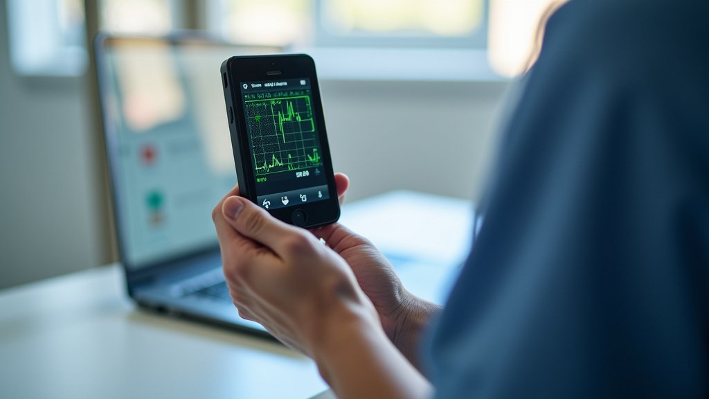
Modern technology has brought ECG monitoring right to your wrist or chest. Many smartwatches now have an ECG feature, and there are adhesive patches you can wear. These devices can often detect an irregular rhythm and prompt you to take an ECG reading. While not always as detailed as a clinical ECG, they are valuable for early detection and monitoring at home. They can alert you to potential issues that you can then discuss with your doctor.
Interpreting the Results: More Than Just Wavy Lines
While the monitor collects the data, a healthcare professional, usually a cardiologist, interprets the ECG tracing. They look at the patterns, measure the intervals between waves, and assess the overall rhythm. It’s like a detective piecing together clues from a complex scene.
What Your Doctor Looks For: A Checklist
When reviewing an ECG for AFib, your doctor will specifically check for:
- Are P waves present and regular?
- Is the R-R interval consistently irregular?
- What is the average heart rate?
- Are there any other unusual wave forms?
- Does the rhythm change over time (if using a Holter)?
This careful analysis helps confirm the diagnosis and guides treatment decisions. It’s a critical step in managing your heart health effectively.
The Importance of Early Detection
Why is all this detection so vital? We found that early diagnosis of AFib is incredibly important for preventing serious complications, especially stroke (CDC). When your atria quiver instead of pumping properly, blood can pool and form clots. If a clot travels to your brain, it can cause a stroke.
By identifying AFib early through ECG monitoring, your doctor can start you on medications or other treatments to manage your rhythm, prevent clots, and significantly reduce your stroke risk. So, if you’re experiencing symptoms like palpitations, shortness of breath, or fatigue, getting an ECG is a very wise step.
Conclusion
Understanding how an ECG monitor detects AFib truly highlights the ingenuity behind these devices. By meticulously analyzing your heart’s electrical signals, they identify the tell-tale signs of chaotic atrial activity and an irregular rhythm. We found that this technology, from simple 12-lead tests to advanced wearable monitors, plays a vital role in protecting your heart health.
Early detection of AFib helps prevent serious complications like stroke. If you’re experiencing symptoms or have concerns about your heart rhythm, talk to your doctor. Getting an ECG could be a simple, life-saving step.
Frequently Asked Questions
Can a normal ECG always rule out AFib?
Not always. A standard 12-lead ECG is a snapshot. If you’re not having an AFib episode during the brief recording, it might appear normal. That’s why your doctor might recommend a Holter or event monitor for intermittent AFib.
Are home ECG devices as accurate as hospital ones?
Home ECG devices, like those in smartwatches or patches, are great for screening and monitoring. They can detect irregularities and prompt you to seek medical attention. However, a doctor-performed 12-lead ECG or longer-term monitor provides more detailed data for a definitive diagnosis.
What is the difference between AFib and other arrhythmias on an ECG?
AFib has specific ECG signatures: no clear P waves and an “irregularly irregular” R-R interval. Other arrhythmias might show different abnormal P wave shapes, consistent but rapid heart rates, or specific extra beats. Your doctor can distinguish these patterns.
Does stress or anxiety affect ECG readings for AFib?
Stress and anxiety can increase your heart rate and even cause palpitations, which can feel similar to AFib symptoms. While they don’t directly cause AFib itself, they can influence your heart rhythm. An ECG can help differentiate between stress-related heart rate changes and true AFib.
How long does it take for an ECG to detect AFib?
A standard 12-lead ECG takes only a few seconds to record. If AFib is present during that brief window, it can be detected immediately. For intermittent AFib, a Holter monitor can detect it over 24-48 hours, or an event recorder over weeks or months.