
How to Interpret AFib on ECG Monitor?
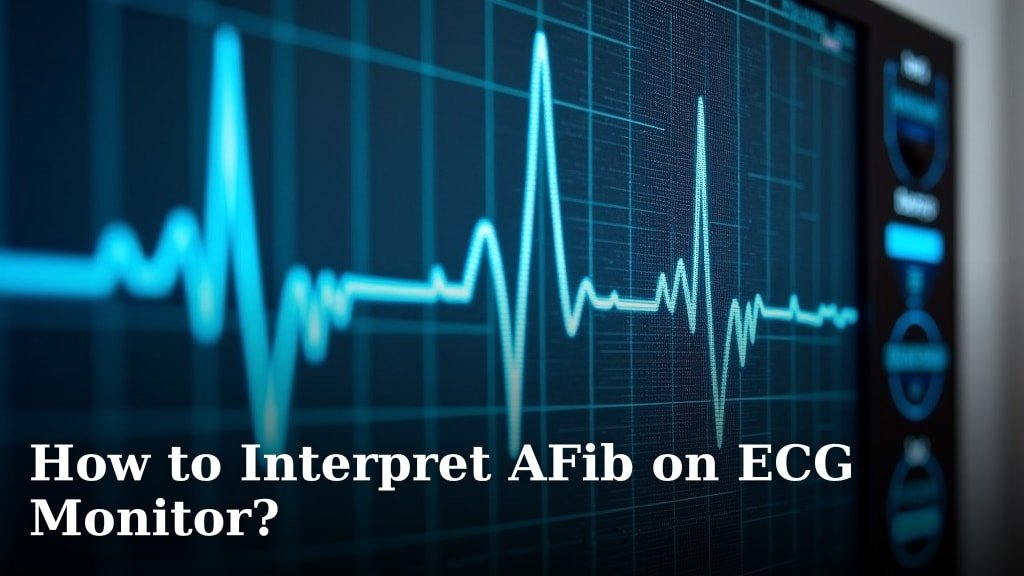
To interpret AFib on an ECG monitor, look for an irregularly irregular heart rhythm along with the absence of distinct P waves. The electrical signals in your heart’s upper chambers are chaotic, not coordinated.
This means your atria are quivering instead of beating properly. You’ll often see small, erratic fibrillatory waves instead of clear, organized P waves before each QRS complex. This irregular electrical activity creates a distinctive pattern on the ECG strip.
- AFib means chaotic atrial electrical activity.
- Look for a heart rhythm with no clear pattern.
- P waves will be missing or look very messy.
- The QRS complexes appear at unpredictable times.
Understanding these key signs can help you recognize Atrial Fibrillation (AFib) on an ECG. Let’s walk through exactly how to spot these changes and what they mean for you.
Understanding AFib on an ECG Strip
Recognizing the signs of AFib on an ECG is simpler than you might think. We’ll break down each key feature, so you know exactly what to look for.
Think of an ECG as a map of your heart’s electrical journey. In AFib, that map gets a little messy.
The Irregularly Irregular Rhythm Explained
One of the most telling signs of AFib is an “irregularly irregular” rhythm. What does that mouthful mean?
Imagine tapping your foot to a song. Normally, there’s a steady beat, right? In AFib, it’s like someone is randomly hitting different drums at different times.
On an ECG, this means the R-R intervals vary without any predictable pattern. The space between each QRS complex (the tall spike) changes constantly.
There’s no rhyme or reason to when the next beat will occur. This lack of pattern is crucial for identifying AFib (American Heart Association).
Comparing Regular vs. Irregular Rhythms
To help you see the difference, let’s look at what a normal rhythm usually shows. You’d see a consistent distance between each beat.
With AFib, those distances are all over the place. Some beats might be close together, others far apart. It’s truly random.
The Missing P Wave: A Key Indicator
Now, let’s talk about P waves. These small bumps usually show your atria contracting properly. They represent the clear electrical signal from your heart’s upper chambers.
In AFib, these organized P waves disappear. Why? Because your atria aren’t contracting in a coordinated way (Mayo Clinic).
Instead of a clear P wave, you’ll often see what we call “fibrillatory waves” or “f waves.” They are small, erratic, and uneven.
These f waves look like a rapid, quivering baseline. They are a jumbled mess of electrical activity, not clean, distinct waves.
Identifying Fibrillatory Waves (f waves)
Look closely at the baseline between the QRS complexes. If you don’t see clear P waves, but instead see these small, wavy irregularities, that’s a strong hint of AFib.
Picture a calm lake surface vs. a choppy, wind-blown pond. The choppy pond represents the f waves.
What About the QRS Complex?
The QRS complex is the most prominent part of an ECG tracing. It shows the electrical activity as your heart’s main pumping chambers (ventricles) contract.
In AFib, the QRS complexes typically look normal in shape and duration. They are usually narrow, just like in a regular rhythm.
The key here isn’t the shape of the QRS, but its timing. Remember the irregularly irregular rhythm we discussed? That applies to the timing of the QRS complexes.
They happen at unpredictable intervals, even though each individual QRS might look fine (Cleveland Clinic).
Normal vs. Aberrant Conduction
Sometimes, very rarely, rapid AFib can cause some QRS complexes to appear wider. This is called “aberrant conduction.”
But for most cases, expect normal-looking QRS complexes that just pop up at random times.
Putting All the Pieces Together
So, you’ve got chaotic atrial activity (no P waves, just f waves) and an irregular ventricular rhythm (QRS complexes at unpredictable intervals). This combination usually points straight to AFib.
A Checklist for Spotting AFib on an ECG
When you’re looking at an ECG strip, here’s a quick mental checklist:
- Is the rhythm completely irregular?
- Are there clear P waves before each QRS? (Hint: in AFib, the answer is no!)
- Do you see small, squiggly f waves instead of P waves?
- Are the QRS complexes generally narrow?
- Is there any pattern to the R-R intervals? (Again, for AFib, no pattern.)
If you answered yes to the first, third, and fourth questions, and no to the second and fifth, you’re likely looking at AFib.
Why is It Irregularly Irregular?
Let’s think about the heart’s natural pacemaker, the SA node. In AFib, the atria are bombarded by thousands of tiny, disorganized electrical impulses, not just the one from the SA node.
It’s like a crowded room where everyone is talking at once, instead of one person speaking clearly.
Only some of these chaotic impulses make it through the AV node (the gatekeeper to the ventricles) to trigger a QRS complex. And they make it through at random times.
This explains the lack of P waves and the unpredictable timing of the QRS complexes.
Understanding Different Types of AFib on ECG
While the core ECG features remain similar, AFib isn’t always a one-size-fits-all diagnosis. Its severity or pattern can sometimes give clues to its type.
Paroxysmal vs. Persistent AFib
You may hear terms like “paroxysmal” or “persistent” AFib. What do these mean for an ECG?
AFib Type
ECG Presentation Clue
Heart Rhythm Behavior
Paroxysmal AFib
AFib signs appear, then disappear.
Starts and stops on its own, usually within 7 days.
Persistent AFib
Consistent AFib for >7 days.
Doesn’t stop on its own; needs intervention to reset.
Long-Standing Persistent AFib
AFib for 1 year or more.
Long-term, continuous AFib.
Permanent AFib
AFib accepted as irreversible.
Decision made not to restore normal rhythm.
On an actual ECG strip, paroxysmal AFib means you might see stretches of normal rhythm that then suddenly switch into the irregularly irregular pattern of AFib.
Persistent AFib, on the other hand, means the AFib pattern is continuous throughout the ECG recording. It’s there for the long haul (NIH).
The ECG recording often just captures a moment in time. Longer monitoring might be needed for full classification.
Rate in AFib: Fast vs. Slow
Another thing to observe is the heart rate during AFib. It can vary quite a bit.
AFib with “rapid ventricular response” (RVR) means your ventricles are beating very fast. The QRS complexes will be frequent and close together, albeit still irregular.
Conversely, AFib with a slower rate means fewer QRS complexes per minute. This might happen if medication is working well, or due to other heart conditions.
The fundamental signs of AFib – no P waves and irregular rhythm – remain, regardless of the overall heart rate.

When to Seek Medical Attention
Spotting AFib on an ECG yourself is a wonderful step towards understanding your health. However, it’s really important to remember that self-interpretation is not a diagnosis.
If you notice these patterns on an ECG monitor, especially if it’s from a home device, contact your doctor promptly. Share your observations and any recorded strips.
You’ll want a healthcare professional to confirm any findings. They can provide an accurate diagnosis, discuss symptoms, and guide you on the best steps forward for your health (CDC).
Remember, the goal is always to keep your heart healthy and happy. Being informed helps you be a good partner in your own care.
Conclusion
You’ve now learned the critical signs of AFib on an ECG. Recognizing the irregularly irregular rhythm, the absence of P waves, and the presence of fibrillatory waves are your key tools. This knowledge helps you understand what’s happening with your heart’s electrical activity. While self-awareness is empowering, remember your role is to observe, not diagnose. If you spot these patterns, share your findings with a healthcare professional immediately for proper evaluation and guidance.
Frequently Asked Questions
Can fitness trackers or smartwatches detect AFib?
Many modern fitness trackers and smartwatches have ECG capabilities. They can detect an irregular heart rhythm that might suggest AFib. However, these devices are screening tools, not diagnostic ones.
Always confirm any findings with a medical-grade ECG and a doctor. It’s a great way to monitor your heart, but professional medical advice is essential.
How often should I check for AFib if I have a home ECG monitor?
The frequency depends on your doctor’s recommendations and your specific health situation. If you have known AFib, your doctor might suggest daily checks or when you feel symptoms. If you’re checking for the first time, follow your device’s instructions, or your doctor’s orders. Don’t over-monitor anxiety can be counterproductive.
What’s the difference between AFib and atrial flutter on an ECG?
Both cause an irregular heart rhythm, but atrial flutter typically shows regular, sawtooth-shaped flutter waves instead of disorganized f waves. AFib’s baseline is chaotic. A healthcare professional can easily distinguish between the two on an ECG.
Does having AFib on an ECG always mean I’ll feel symptoms?
Not necessarily. Many people with AFib report no symptoms at all. This is called “silent AFib.” The ECG can still show the characteristic chaotic electrical activity. This is why routine checks or incidental findings are important for proper diagnosis and management, even without symptoms.
If my ECG shows AFib, what’s the next step my doctor might take?
Your doctor will likely order a 12-lead ECG for a more comprehensive view and might suggest a Holter monitor for 24-48 hours to capture intermittent episodes. They will also review your medical history and discuss potential treatment options. These can include medications, lifestyle changes, or procedures to manage your heart rhythm.