Interpreting EKG Lines: What the Squiggles Mean
EKG lines show your heart’s electrical activity through specific wave patterns that represent different phases of your heartbeat.
Each squiggle on the EKG strip has meaning – P waves show atrial activity, QRS complexes show ventricular contractions, and T waves show recovery phases.
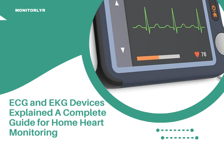
You’re staring at that squiggly line on the EKG monitor, and it looks like random scratches. But those “squiggles” tell an amazing story about your heart’s electrical system.
Think of your heart as a house with its own electrical wiring. Every beat starts with an electrical signal that travels through specific pathways. The EKG captures this journey and translates it into waves you can see.
The Basic EKG Wave Pattern
Your normal heartbeat creates a predictable pattern on the EKG. This pattern repeats with every heartbeat, like a signature your heart writes over and over.
The main components are waves, segments, and intervals. Each piece gives doctors different information about how your heart works.
P Wave: The Starting Signal
The P wave is that small bump before the big spike. It shows your atria (the top chambers) getting ready to squeeze.
A normal P wave is smooth and rounded. It should be upright in most leads. If it’s too tall, too wide, or missing, that tells doctors something about your atrial function.
What P Wave Problems Mean
When P waves look abnormal, it often points to atrial issues. Tall P waves might mean your right atrium is working too hard. Wide P waves could suggest left atrial problems.
Missing P waves? That’s a big clue. Your heart might be beating from a different location than normal.
QRS Complex: The Main Event
That big spike you notice first? That’s the QRS complex. It represents your ventricles (the main pumping chambers) contracting.
The QRS should be narrow and sharp. It’s usually the tallest part of the EKG because your ventricles are your heart’s strongest muscles.
QRS Width Matters
A normal QRS lasts less than 0.12 seconds. When it’s wider, the electrical signal is taking a detour. This could mean blockages in your heart’s electrical pathways.
Wide QRS complexes often point to bundle branch blocks or other conduction problems. Think of it like traffic taking side streets when the highway is blocked.
QRS Height and Shape
Very tall QRS complexes might suggest your ventricles have grown thicker. This happens when your heart works harder than normal for long periods.
The shape matters too. Smooth, pointed peaks are normal. Jagged or notched patterns can indicate different problems.
T Wave: The Recovery Phase
The T wave comes after the QRS complex. It shows your ventricles resetting and getting ready for the next beat.
Normal T waves are asymmetrical – they slope up gradually and come down more steeply. They should point in the same direction as your QRS complex in most leads.
T Wave Abnormalities
Flipped T waves can signal heart muscle problems. Very tall, peaked T waves might indicate high potassium levels. Flat T waves could suggest low potassium or other issues.
Changes in T waves often develop gradually. They can be early signs of heart muscle stress or damage.
ST Segment: The Flat Line
Between the QRS complex and T wave lies the ST segment. It should be flat and level with the baseline.
When the ST segment rises or falls significantly, it’s a major red flag. ST elevation often means active heart attack. ST depression can indicate reduced blood flow to the heart muscle.
Why ST Changes Matter
The ST segment represents the time when your ventricles are fully contracted. During this phase, the electrical activity should be quiet.
Any deviation from the flat baseline suggests the heart muscle isn’t getting enough oxygen or is actively being damaged.
Heart Rhythm Patterns
Beyond individual waves, the overall pattern tells you about heart rhythm. Regular spacing between beats indicates normal sinus rhythm.
Irregular patterns can mean different types of arrhythmias. Some are harmless. Others need immediate attention.
Rate and Regularity
Normal heart rates range from 60 to 100 beats per minute at rest. You can calculate this by counting QRS complexes over a specific time period.
Regularity means consistent spacing between beats. Your heart should beat like a steady drum, not like random raindrops.
Common Rhythm Problems
Atrial fibrillation shows up as an irregularly irregular pattern with absent P waves. The baseline might look wavy instead of smooth.
Heart blocks show up as disconnected P waves and QRS complexes. Sometimes P waves appear without following QRS complexes.
Reading EKG Leads
Standard EKGs use 12 different views called leads. Each lead looks at your heart from a different angle, like taking photos from different positions.
The same heart problem might look different in different leads. That’s why doctors examine all leads together.
Lead Groups
Limb leads (I, II, III, aVR, aVL, aVF) show your heart’s electrical activity in the frontal plane. Chest leads (V1-V6) show the horizontal plane.
Different leads highlight different areas of the heart. Changes in specific lead groups help pinpoint problem locations.
Common EKG Findings
| Finding | What It Looks Like | Possible Meaning |
|---|---|---|
| Normal Sinus Rhythm | Regular, identical beats | Healthy heart function |
| Sinus Tachycardia | Fast but regular rhythm | Exercise, stress, fever |
| Sinus Bradycardia | Slow but regular rhythm | Athletic heart, medications |
| Atrial Fibrillation | Irregular, no clear P waves | Atrial rhythm disorder |
When to Worry
Some EKG changes need immediate attention. ST elevation, new wide QRS complexes, or very fast irregular rhythms are emergency findings.
Other changes develop slowly over time. These might indicate chronic conditions that need monitoring and treatment.
Factors That Affect EKG Readings
Your EKG can change based on many factors. Body position, breathing, medications, and electrode placement all make differences.
Even your body build affects the EKG appearance. Thin people often have taller waves. Larger people might have smaller waves.
Technical Issues
Poor electrode contact creates artifact that looks like extra squiggles. Movement during recording can make the baseline wavy.
Always tell your technician if you feel uncomfortable or need to move during the test.
Conclusion
Those squiggly lines on your EKG tell a detailed story about your heart’s electrical system. Each wave, segment, and interval provides specific information about different aspects of your heart function. While the patterns might look random at first glance, they follow predictable rules that trained professionals can read like a book. Understanding the basics helps you appreciate the amazing technology that lets us see your heartbeat in action. Remember that EKG interpretation requires medical training, so always discuss your results with qualified healthcare providers who can put the patterns into context with your overall health picture.
Can I learn to read my own EKG results?
You can learn basic EKG concepts, but accurate interpretation requires extensive medical training. Focus on understanding general patterns rather than trying to diagnose specific conditions from your EKG strips.
Why do different EKG machines show slightly different results?
Machine calibration, electrode placement, and filtering settings can create minor variations between devices. These differences are usually insignificant for clinical interpretation, but they explain why your EKGs might look slightly different at different facilities.
How long does heart rhythm need to be monitored to get accurate results?
A standard 12-lead EKG captures about 10 seconds of rhythm, which is enough for most basic assessments. However, intermittent problems might require longer monitoring periods using Holter monitors or event recorders that track your rhythm for days or weeks.
Can anxiety or stress change my EKG appearance?
Yes, anxiety and stress can increase your heart rate and sometimes create minor ST segment changes. These are usually temporary and resolve once you relax, but persistent stress-related changes should be evaluated by your doctor.
Do normal EKG results guarantee my heart is healthy?
A normal EKG is reassuring, but it only shows electrical activity during those few seconds of recording. Some heart conditions don’t always show up on resting EKGs, which is why doctors might recommend stress tests, echocardiograms, or other cardiac evaluations based on your symptoms and risk factors.